Product Description
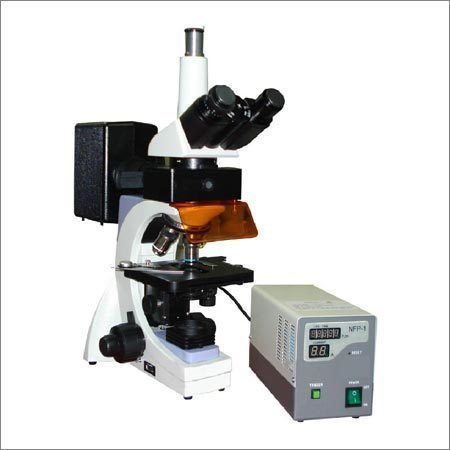
To meet the growing demands of the medical industry we offer superior quality Co-Axial Trinocular Research Microscope with micro-photographic system. Our designed with our cutting edge technology our laboratory microscopes are provided with:
- Fine focusing system and latest co-axial coarse based on a 4-gear reduction system
- Traveling on ball bearing guides with highly sensitive fine motion
- Graduation reading to 0.002 mm.
- Large graduate mechanical stage (145 x 125mm.
- Convenient slide manipulation.
- Large graduate mechanical stage (145 x 125mm)
- Convenient slide manipulation.
Optical Combination:
Some of the features are enlisted below:
Online Respiratory Disturbance Index (R.D.I.) Calculation.
| Ideally suitable in ICU's conditions. |
| Complete system Windows ME/XP. |
| USB connectivity, no external power supply required. |
| Online sleep stage scoring. |
| Tabular format for all important readings. |
| Graphical & Numerical Analysis. |
| Facility to change individual channel HF, LF, Sensitivity. |
| Optical isolated to provide excellent S/N performance. |
It is provided with Automatic Marking :
| Apneas & Hypopneas: (obstructive central or mixed apneas). |
| Sleep stage 1, 2, 3, & 4. |
| Arousale REM. |
Following Reports are printed after the test is completed :
| Oxygen De saturation, HR Variation, Body Position. |
| Limp movement, snoring, summary report. |
| Sleep stages: S1, S2, S3, S4, REM, Wake time. |
| Auto test start from same stage on power resumption. |
| Selectable sweep speed for careful data observation. |
| Split Screen, freeze facility to study event carefully. |
| Highly synchronize video & PSG data |
| Online display of heart rate, RDI & sleep stages. |
| Ultra fast automatic paging facility for efficient analysis. |
| Archiving on any media. |
| High resolution auto focus camera with remote control. |
| Patient snap photo on PSG report for easy identification |
| Off line room Audio playback |
| Complete patient questionnaire before and after sleep study |
Provided with maintenance accessories and packed in a thermocole and wooden box.
Superior Optical Precision & FlexibilityWith its high-quality achromatic objectives-4x, 10x, 40x, and 100x oil immersion-and standard 160 mm optical tube, this microscope delivers crisp images at magnifications from 40x to 1000x. The monocular head is 45 inclined and 360 rotatable, providing ergonomic viewing for students and professionals alike.
Built for Durability and StabilityConstructed from die-cast mild steel and finished with a smooth, powder-coated surface, this microscope withstands daily use and resists corrosion. The rugged, anti-slip base and curved, rigid arm ensure vibration-free operation and easy portability within the laboratory environment.
Practical Accessories and Easy MaintenanceEssential accessories such as a dust cover, immersion oil, and cleaning cloth are included, supporting longevity and ease of maintenance. Spare parts, including bulbs (for lamp versions) and extra eyepieces, are readily available, ensuring years of reliable use.
FAQ's of Laboratory Microscope :
Q: How do I move a microscope slide with manual X-Y stage movement?
A: To move your slide, use the manual X-Y adjustment knobs on the stage, allowing precise lateral and longitudinal positioning for accurate specimen examination.
Q: What are the key benefits of the microscope's powder-coated, anti-fungal finish?
A: The smooth powder-coated paint protects the frame from corrosion and the anti-fungal coating prevents microbial growth, ensuring long-lasting performance even in humid laboratory environments.
Q: Where is this microscope best suited for use?
A: This microscope is ideal for indoor environments such as laboratories, educational institutions, and research centers due to its robust build and optical precision.
Q: When should I use immersion oil with this microscope?
A: Immersion oil should be used when working with the 100x oil immersion objective to enhance image clarity and resolution by reducing light refraction between the slide and the lens.
Q: What is the process for focusing on a specimen with this microscope?
A: Begin by using the coarse adjustment knob to get a general focus, then refine the image using the fine adjustment knob, which allows for precise focusing down to 0.002mm per division.
Q: How is the microscope illuminated during use?
A: The primary light source is the plano-concave mirror, which redirects natural or external light to illuminate the specimen. For increased flexibility, an optional substage lamp may be requested.


 English
 Spanish
 French
 German
 Italian
 Chinese (Simplified)
 Japanese
 Korean
 Arabic
 Portuguese