Product Description
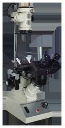
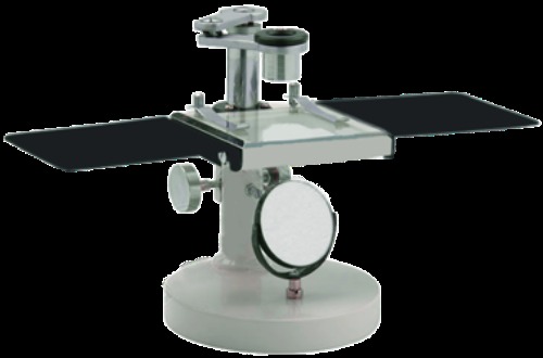
The Dissecting Microscope is used for demonstration, close study of small organisms and fine dissection work. Dissecting Microscope high-eyepoint lenses 10x & 20x have alternative use on a jointed arm allowing the whole stage area to be scanned. Dissecting Microscope stage 85mm X 75mm provided with spring stage clips, has a Glass Plate with arrangement for inserting white/black metal plate for respective background.
Substage plano-concave mirror reflects sufficient daylight & artificial light on the object. Dissecting Microscope Rack & Pinion mechanism is enclosed within the stage support pillar and operated by a knob on either side. Detachable hand rests on both sides of the stage provide for a steadying support during dissection.
Base - Heavy round base with precisely designed body.
Focussing - Sensitive focussing, done by rack and pinion arrangement. Revolving arm provided for moving magnifying lens over full stage area.
Stage - 85mm x 75mm with glass plate.
Illumination - A plano-concave lens fitted in fork for light reflection.
Optics - 10x & 20x Eyepieces
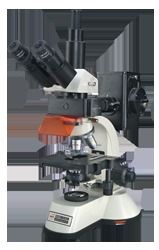
Sturdy Build for Reliable PerformanceCrafted with sturdy, mild steel and finished in durable, chemically resistant paint, this dissecting microscope is built to withstand continuous use in educational and laboratory environments. The heavy cast iron base ensures minimal vibration during operation, guaranteeing clearer, more stable observations. This robust construction supports long-term reliability and safe handling.
Flexible Observation and Simple OperationThe microscope features a 360 rotating arm and a monocular, 45 inclined tube, allowing users to adjust viewing angles easily for comfortable dissection and study. Coarse focusing is accomplished via the rack and pinion mechanism with an adjustment range of up to 25 mm. With interchangeable stage plates (glass and opaque), it accommodates various specimen types, making it adaptable and user-friendly.
Efficient Illumination and Easy MaintenanceIllumination is achieved using a sub-stage plano-concave mirror, optimizing the use of ambient or external light sources for clear visualization. Anti-fungal coatings on optical elements and a scratch-resistant body finish ensure longevity and hygiene. Cleaning and general maintenance are simple, making this microscope ideal for routine educational and laboratory dissection work.
FAQ's of Dissecting Microscope:
Q: How do I operate the dissecting microscope for routine laboratory work?
A: Place the specimen on the stage plate and secure it with the metal clips. Adjust the viewing angle using the 360 rotating arm and the inclined monocular tube. Select the appropriate eyepiece (10x or 20x) and objective, then focus using the coarse rack and pinion system. Use the sub-stage mirror to direct natural or external light onto the specimen for clearer observation.
Q: What maintenance steps are recommended for optimal performance?
A: Clean the lenses gently with a soft, lint-free cloth and avoid harsh chemicals. The anti-fungal coating helps protect optics, but keep the microscope covered with the included dust cover when not in use. Periodically check and tighten stage clips and ensure the mirror is clean for best illumination results.
Q: When should I use the glass versus the opaque stage plate?
A: Use the glass stage plate for transparent or semi-transparent specimens where transmitted light enhances visibility. The opaque stage plate is suitable for opaque specimens, providing contrast and better surface detail under reflected light.
Q: Where is this microscope best utilized?
A: This dissecting microscope is ideal for school and college biology labs, and for routine laboratory dissections where robustness, portability, and straightforward manual operation are required. Its simple, compact design and durable construction suit both educational and professional settings.
Q: What are the key benefits of using this microscope in educational settings?
A: Its sturdy build ensures longevity, while the easy-to-use focusing system and flexible head movement support quick setup and comfortable viewing. The anti-fungal coated optics and scratch-resistant body ensure hygiene and durability, making it an excellent investment for teaching and training purposes.
Q: How does the magnification system work on this microscope?
A: Magnification is achieved using pairs of interchangeable wide field eyepieces (10x and 20x) in combination with the fixed or rotatable 2x achromatic objective. The optical resolution depends on the chosen eyepiece and objective pairing, providing either 10x or 20x total magnification for visual/manual observation.


 English
 Spanish
 French
 German
 Italian
 Chinese (Simplified)
 Japanese
 Korean
 Arabic
 Portuguese