Product Description
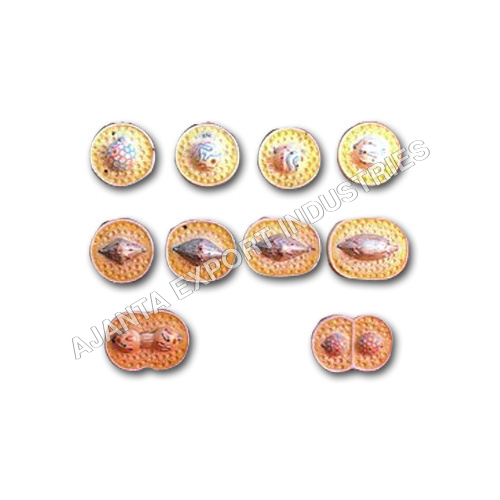
Discover the brilliance of cellular science with our New Animal Cell Division Mitosis Model, now on sale-a true bargain for students and educators alike. This classic educational tool superbly represents every inestimable stage of mitosis, including Prophase, Metaphase, Anaphase, Telophase, and Cytokinesis. Crafted from high-quality, color-coded plastic, the model features hand-painted, removable parts for brilliant teacher demonstrations. With a glossy, easy-to-clean finish and a stable base stand, it's perfect for biology classrooms, labs, and medical settings. English stage labels and a comprehensive manual ensure accurate, clear learning for ages 11 and above.
Comprehensive Application & User Profiles
The Animal Cell Division Mitosis Model is ideal for enriching biology study sessions in schools, colleges, and medical institutes. Teachers, students, and laboratory professionals use it for hands-on demonstrations and research references. Its manual, three-dimensional construct facilitates interactive learning, making complex cellular divisions easy to understand. This model's versatility allows it to be used in classrooms, medical labs, and even hospitals, serving users aged 11 years and older with detailed accuracy.
Payment, Export Markets & Packaging Highlights
Our mitosis models are shipped worldwide, reaching main export markets such as North America, Europe, and Asia. We offer flexible payment terms and ensure each model is carefully packaged to prevent transit damage. The outlay includes protective wrapping and sturdy boxes, enabling safe delivery to distributors, exporters, and retailers. Rest assured, every order is processed promptly, and models arrive ready for display with all included components and guides.
FAQ's of Animal Cell Division Mitosis:
Q: How is the Animal Cell Division Mitosis Model used in educational settings?
A: This model is widely used as a visual aid during classroom lectures, teacher demonstrations, and laboratory sessions to illustrate and explain the stages of mitosis within an animal cell.
Q: What stages of mitosis are represented in the model?
A: The model accurately depicts all key stages of mitosis, including Prophase, Metaphase, Anaphase, Telophase, and Cytokinesis, making cellular division easy to visualize and study.
Q: Where can this mitosis model be applied outside the classroom?
A: Besides educational institutions, the model is also suitable for use in medical laboratories and hospitals for staff training and patient education on cell biology processes.
Q: What benefits does the three-dimensional, hand-painted design offer?
A: The detailed, three-dimensional, hand-painted design enhances engagement and learning, allowing users to observe cell components from multiple angles and better understand their arrangement during mitosis.
Q: How should the model be maintained for long-lasting use?
A: Maintaining this model is simple-just wipe its glossy, easy-to-clean surface with a damp cloth to keep it in pristine condition for repeated educational use.


 English
 Spanish
 French
 German
 Italian
 Chinese (Simplified)
 Japanese
 Korean
 Arabic
 Portuguese