Product Description
ProductDescription
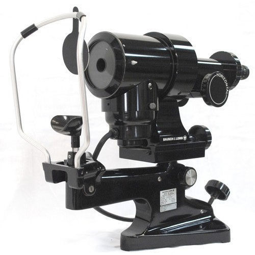
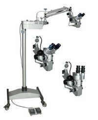
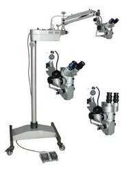
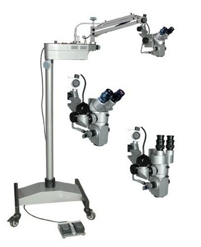
SURGICAL MICROSCOPE 180Degree Inclinable BinocularsLED Illumination, with Image Display System
Technical Specifications:
BINOCULARS:-
Binocular Tubes :0 to 180 deg. Inclinable Binoculars
MAGNIFICATION SYSTEM:-
Magnifications : 5 Step [ 4x, 6x, 10x, 16x &25x ]
EYE PIECES (OPTIONAL)
Eye Pieces : 12.5x
MAGNIFICATION RANGES:-
Objective Lens : F=200mm
Field of View : 50mm
IPD Adjustment : 55mm to 75mm
FOCUSING:-
Fine Focusing : Manual Fine Focusing by Knob
ILLUMINATION:-
Illumination : 100W LED Lamps with 100% coaxial through thelenses focused Fiber optics light guide 1.5 meter, bright WHITE LED light,maximum at standard condition
Field of Illumination : 70mm
Arm : Counter balanced pantographic arm with320 degree rotation.
Vertical Range of Counter BalancedArm : 250mmto 300mm
Floor Stand : Mobile floor stand on caster wheels foreasy handling and absolute stability.
Base : 600mm x 500mm (Approx)
Height : 1500mm (Approx.)
Weight : 65kg (Approx.)
Power : AC 220V, 50/60Hz. (110V on request)
Innovative Magnification and ErgonomicsExperience precise surgical visualization with a variable magnification range from 4x to 40x and 10x widefield eyepieces. The binocular head offers ergonomic adjustment and a full 180 rotation, ensuring comfort and optimal viewing angles during long procedures. Customizable working distances between 200 mm to 400 mm accommodate diverse surgical requirements.
Advanced Illumination and ImagingDual LED light sources provide uniform illumination, while integrated yellow, green, and IR filters enhance tissue differentiation and visualization. The built-in photo/video port with C-mount expands documentation options, making this microscope ideal for both clinical work and teaching. Anti-reflective coatings and apochromatic, multi-layer lenses maximize resolution and image clarity.
User-Friendly Operation and MobilityAn articulated arm with 4-way movement and a touch pad based control panel allow intuitive microscope positioning. Floor stand with castors and integrated anti-vibration system ensures smooth, stable operation and easy transport within the operating room. Sterilized external surfaces and wipe-down sterilization mode support strict hygiene standards.
FAQ's of Surgical Microscope:
Q: How do I adjust the magnification on the surgical microscope?
A: You can vary the magnification stepwise between 4x and 40x using the microscope's control panel, ensuring optimal visualization according to procedural needs. The apochromatic lenses ensure high resolution at all magnification levels.
Q: What processes are involved in sterilizing the surgical microscope?
A: Sterilization is performed by wiping down the external surfaces, which are designed to be easily cleaned. The microscope features medical grade material and anti-reflective coating, ensuring compliance with surgical hygiene standards.
Q: When is this surgical microscope typically used?
A: This microscope is utilized in ophthalmic, neurosurgery, ENT, and dental surgeries where high precision and magnified visualization are necessary for delicate procedures.
Q: Where should the microscope be positioned during surgery?
A: The floor stand with castors allows flexible positioning near the surgical site. The articulated arm provides four-way movement for tailored adjustment, maximizing surgical access without compromising stability due to the integrated anti-vibration system.
Q: What are the main benefits of the motorized fine and coarse focus adjustment?
A: Motorized focus enables surgeons to make swift, accurate adjustments during operations, maintaining image clarity and reducing manual strain. This is particularly important during lengthy and complex procedures.
Q: How does the built-in photo/video port enhance documentation?
A: The integrated photo/video port and C-mount allow real-time recording and imaging of surgical procedures, beneficial for medical documentation, case studies, and educational purposes.
Q: What filter options are available, and how do they improve surgical outcomes?
A: The microscope includes integrated yellow, green, and IR filters that improve contrast and tissue visualization. This helps surgeons differentiate anatomical structures more clearly and operate more safely.


 English
 Spanish
 French
 German
 Italian
 Chinese (Simplified)
 Japanese
 Korean
 Arabic
 Portuguese