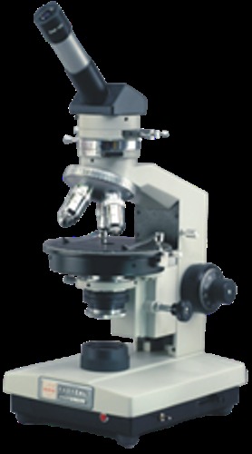
Product Description
Research Polarizing Microscope are principal piece of equipment used by the Geologists to observe the optical properties of minerals. It has indispensable applications for petrography, crystallography, geology, mineralogy, ceramics, toxicology, chemical microscopy or detecting defects in semi-conductors, finding stress points in metals, glass & textiles etc.
Stage - Compact Ball bearing Rotating Stage, 140mm dia., graduation with vernier, lock & stage specimen clips.
Focusing - Coaxial focusing is provided with coarse tension adjustment ring and graduated slow motion knobs. Adjustable safety lock is attached to prevent the breakage of specimen slides.
Illuminator - Koehler's Illuminator with pre-centered 20W Tungsten Halogen source & integral electric transformer with intensity control. Input Voltage 220 or 110V.
Intermediate Tube - Built-in focusable Bertrand Lens with center adjustment. Built-in Analyzer removable from optical part. Analyzer can be rotated 90º with vernier reading to 6'. Compensator 1 Lambda, 1/4 Lambda & Quartz Wedge can be inserted in the tube as per requirement.
Compensator - Gypsum (1st. order red compensating plate, 560nm). Mica (145nm) & Quartz Wedge (I-IV order).
Objectives - Stress less flat achromatic DIN Objectives PF4x/0.10, PF10x/0.25 & PF40x/0.65 SL
Eyepieces - HKW10x Paired & WF 10x Cross
Polarizer - Fully rotatable Polarizer on a strain free N.A 1.25 abbe condenser with iris diaphragm.
Optional Accessories - Detachable Mechanical Stage, Objectives PF20x & PF60X, Micrometer Eyepiece WF10x, Microphotographic Equipment, 30º Inclined Siedentopf Heads. Wooden storing cabinet etc.
Engineered for Hospital ResearchOur Research Polarizing Microscope is specifically tailored for hospital laboratories, enabling precise examination of crystals, minerals, and biological tissues. Its sturdy stainless steel build ensures long-term reliability in demanding environments, while manual controls offer exceptional user control during studies. By integrating laser technology, users benefit from high-resolution optics essential for rigorous medical research.
Precision and Clarity through Laser TechnologyFeaturing advanced laser-assisted optics, this microscope delivers heightened contrast and sharpness when analyzing birefringent materials. This technological edge allows for detailed visualization, catering to the exacting needs of modern hospital research. Medical experts can trust its accuracy for diagnostic assessments or research applications, contributing to better patient outcomes.
FAQ's of Research Polarizing Microscope:
Q: How does the Research Polarizing Microscope benefit hospital laboratories?
A: The Research Polarizing Microscope enhances hospital laboratories by providing high-precision analysis of complex materials, such as tissues and crystalline structures. Its advanced optics and polarization capabilities support accurate diagnostics and research, ultimately improving laboratory workflow and patient care.
Q: What materials is the Research Polarizing Microscope made from, and why is this important?
A: This microscope is made from high-quality stainless steel, which offers strength, corrosion resistance, and easy cleaning. This makes the unit highly durable and suitable for repeated use in hospital environments, ensuring longevity and consistent performance.
Q: When should a hospital consider using a research polarizing microscope?
A: Hospitals should utilize a research polarizing microscope when in-depth analysis of anisotropic (birefringent) materials is required. It is particularly useful for laboratories conducting mineralogy, pathology, or research involving crystal structures or specialized tissue studies.
Q: Where can I obtain this Research Polarizing Microscope in India?
A: The Research Polarizing Microscope is available through reputable distributors, exporters, manufacturers, suppliers, and traders across India. You can contact established medical equipment companies or authorized microscope providers to acquire this equipment.
Q: What is the process for using the manual driven method on this microscope?
A: Using the manual driven method involves adjusting the stage, focus, and polarization filters by hand for precise alignment and observation. This method gives the user direct control over the viewing process, allowing for meticulous examination of samples throughout research sessions.
Q: How does the integration of laser technology improve the microscope's performance?
A: Laser technology integrated within this microscope enhances image clarity and contrast, making it easier to differentiate between materials with similar properties. This leads to more accurate observations and improved research results, especially for complex medical or scientific analyses.


 English
 Spanish
 French
 German
 Italian
 Chinese (Simplified)
 Japanese
 Korean
 Arabic
 Portuguese