Product Description
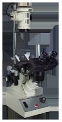
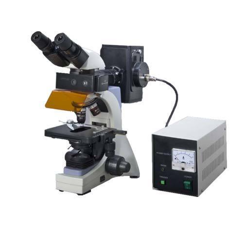
Head: gemel type of trinocular head, inclined at 45 degre
- Optical System: Innity chromatic correcnce Plan Objectives, 4X (0.10), 10x (0.25),
- 40x (0.65),100x (1.25) "
- Phase Contrast Plug-in Plate: Adjustable phase center - 10X/20X, 40X, bright eld;
- phase center presetting - 10X/20X, 40X, bright tion
- "Objective: 1) Innity Corrected Fluoresceeld (optional)
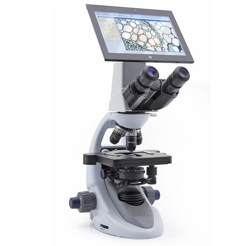
Exceptional Fluorescence ImagingAchieve vivid visualization of fluorescently labeled samples with advanced filter sets for FITC, TRITC, DAPI, and GFP. The high-intensity illumination and precision optical system provide high-contrast, sharp images, vital for research, diagnostics, and teaching applications.
Robust, Ergonomic DesignConstructed with a sturdy die-cast metal frame and anti-mold optics, this microscope endures rigorous laboratory use. The double-layer mechanical stage and trinocular head, coupled with ergonomic controls, ensure comfortable operation even during extended sessions.
Integrated Digital ImagingOptional high-sensitivity CMOS camera modules enable the capture of crisp still images and Full HD videos at up to 30 fps. USB and HDMI output interfaces provide flexible connectivity, allowing seamless documentation, analysis, and sharing of results.
FAQs of Flurorescent Research Microscope:
Q: How does the fluorescence filter system enhance research capabilities?
A: The four-position filter turret, equipped with sets for FITC, TRITC, DAPI, and GFP, allows researchers to swiftly switch between different fluorescence channels. This enhances the ability to study multiple targets within the same sample without changing microscopes, thereby improving workflow and experimental flexibility.
Q: What are the benefits of the infinity-corrected optical system and plan achromatic objectives?
A: Infinity-corrected optics provide consistent image quality across a wide field of view, minimizing optical aberrations. The plan achromatic anti-fungal objectives deliver flat, sharp images with accurate color rendition, essential for precise observation and documentation of fluorescent specimens.
Q: Where can this microscope be effectively utilized?
A: This system is ideally suited for research laboratories, clinical diagnostics, universities, and industrial settings. Its robust construction and high-precision optics make it ideal for cell biology, microbiology, pathology, and materials sciences applications.
Q: How is stage movement controlled during sample examination?
A: The microscope features a graduated double-layer mechanical stage with travel range of 75 x 50 mm and user-friendly controls, allowing precise and repeatable movement across the specimen for accurate observation and imaging.
Q: When is it advantageous to use the trinocular observation head and camera mount?
A: The trinocular head facilitates simultaneous viewing and digital imaging. Its particularly beneficial during collaborative research, teaching sessions, or when recording and analyzing image data using the C-mount-compatible CMOS camera module.
Q: What are the steps to capture images and videos using the integrated camera system?
A: Attach the optional CMOS camera to the C-mount adapter, connect via USB or HDMI, and use the provided software to view, capture, and save images or videos in popular formats like JPEG, BMP, or PNG. This process simplifies documentation and enables efficient data management.
Q: What measures ensure mechanical and optical stability during high-magnification work?
A: Height-adjustable rubber anti-vibration feet and a heavy die-cast frame reduce external vibrations, while the coaxial coarse and fine focus systems with tension adjustment ensure smooth and stable focusing, even at high magnification levels.


 English
 Spanish
 French
 German
 Italian
 Chinese (Simplified)
 Japanese
 Korean
 Arabic
 Portuguese