Product Description
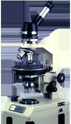
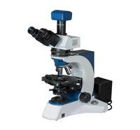
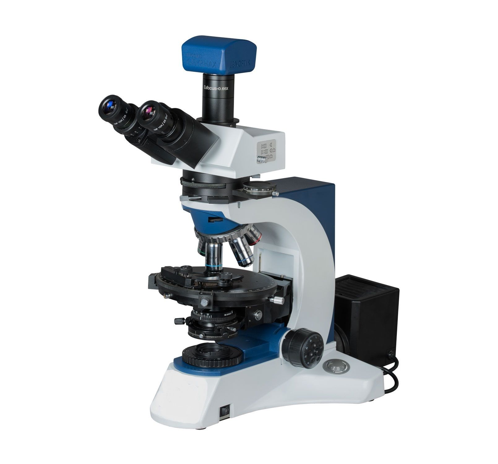
Pol microscopes produce brighter, clearer, and higher contrast images. Facilities for diascopic and episcopic microscope illumination makes it a sought after microscope petrological, geological etc labs. The Bertrand lens is also focusable and centerable.Precision and Flexibility in Polarising MicroscopyFeaturing a 360 rotatable analyzer with vernier scale, the microscope delivers exceptional accuracy for measuring angular changes in specimens. The heavy-duty base and anti-vibration construction offer stability, essential for research-grade analyses. With a 160mm graduated stage and reliable X-Y movement, users can achieve precise positioning and microscale measurements for a range of samples including minerals, crystals, and polymers.
Enhanced Observation CapabilitiesEquipped with a centrable built-in Bertrand lens, the microscope supports both conoscopic and orthoscopic observations, pivotal in crystallography and optical mineralogy. The slot for accessory compensators facilitates advanced study of birefringence using gypsum, mica, or quartz wedge plates. Wide field eyepieces with polarising cross hair, trinocular viewing head, and phototube enable simultaneous visual and digital documentation.
Ergonomic and Durable Design for Research EnvironmentsThe microscope is engineered for extended laboratory usage, featuring an ergonomic, inclined trinocular head and corrosion-resistant body. Adjustable illumination with halogen or LED modules, comprehensive mechanical and optical interfaces, and a wide range of included accessories make it an optimal choice for demanding research applications. Its anti-fungus coated objectives ensure longevity even under high humidity conditions.
FAQs of ADVANCED RESEARCH POLARISING MICROSCOPE:
Q: How does the rotatable analyzer with vernier scale improve measurement accuracy in this polarising microscope?
A: The 360 rotatable analyzer, coupled with the precise vernier scale (reading up to 0.1), allows researchers to measure and analyze specimen polarization angles and optical properties with high accuracy, supporting advanced quantitative studies in mineralogy and material science.
Q: What types of specimens or research applications is this microscope suitable for?
A: This microscope is ideal for polarised light analysis of geological samples, minerals, crystals, polymers, and anisotropic materials. It supports both transmitted and reflected light studies, making it suitable for a wide range of research in geology, chemistry, and material sciences.
Q: Where is the microscope best used, and what operating conditions are recommended?
A: It is best used in research laboratories, universities, and specialized analysis centers. Recommended ambient conditions are temperatures between 1040C and humidity 65% RH, in a non-condensing environment to maintain optimal performance and longevity.
Q: What is the process for attaching a digital camera to the microscope for imaging?
A: The microscope features a trinocular 360 rotatable drawtube and a dedicated phototube. Simply connect a compatible digital camera to the phototube port as per the user manual instructions. This enables direct optical imaging or video recording of observed specimens.
Q: How does the mechanical X-Y stage facilitate sample examination?
A: The stage offers high-precision movement (approx. 75mm x 45mm) with vernier scales for accurate sample positioning and angular measurements. The 160mm diameter rotatable stage has graduated markings and click stops, streamlining alignment and repeated observations during advanced studies.
Q: What are the benefits of strain-free optics and anti-fungus coated objectives?
A: Strain-free optics and anti-fungus coated objectives minimize optical distortions and protect lenses from fungal growth, ensuring clear, reliable imaging and extended durabilityespecially important in moist laboratory environments or for prolonged research use.
Q: When should accessory compensators like gypsum or mica plates be used, and what do they offer?
A: Use compensator plates when studying birefringence and optical retardation in specimens. They help enhance contrast, measure optical path differences, and support advanced qualitative and quantitative analysis in polarised light microscopy.


 English
 Spanish
 French
 German
 Italian
 Chinese (Simplified)
 Japanese
 Korean
 Arabic
 Portuguese